
Do Peptides Cross the Blood-Brain Barrier? What the Evidence Actually Shows
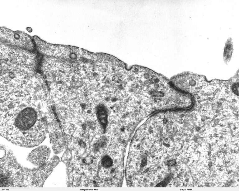
Very few do, and most claims that they do rest on the wrong experiment. The barrier defeats peptides through tight junctions, minimal pinocytosis and active efflux; the routes that are genuinely demonstrated in vivo are receptor-mediated transcytosis vectors, not the compounds usually marketed on BBB penetration.
Most nootropic peptide marketing rests on a premise it never states: that the molecule reaches the brain. For peptides, that premise is the hardest part of the entire pharmacological argument to establish. A compound can be stable in plasma, bind its target at nanomolar affinity and behave beautifully in cultured neurons while still failing at the single step that decides whether any of it matters in a whole animal. What follows is what the barrier does, which routes past it are demonstrated, and how far the usual evidence falls short of the claim it supports. Every compound named is a reference material supplied strictly for laboratory research use only (RUO); nothing below is dosing guidance, a therapeutic claim or a suggestion of human use.
What is the barrier, mechanically?
The blood-brain barrier is not a membrane draped over the brain; it is a property of the cerebral capillaries themselves. Brain endothelial cells are continuous and non-fenestrated — lacking the pores that let solutes leak out of capillaries almost everywhere else — and are sealed to one another by tight junctions of claudin-5 and occludin, anchored to the cytoskeleton by zonula occludens proteins such as ZO-1.1 That seal closes the paracellular route, which for a hydrophilic peptide is the obvious way through.
The second feature matters more than it is usually credited: brain endothelium has an unusually low rate of pinocytosis. Peripheral capillaries move material in bulk by vesicle traffic; brain capillaries largely do not, closing the non-specific vesicular route as well. These cells sit within a neurovascular unit — pericytes embedded in the basement membrane at a far higher ratio than in peripheral vessels, astrocyte end-feet ensheathing the vessel — which induces and maintains the barrier phenotype rather than forming a second wall.1
The third feature is active, and it defeats most candidate molecules. The luminal membrane is densely populated with ATP-driven efflux transporters, principally P-glycoprotein (ABCB1) and breast cancer resistance protein (BCRP/ABCG2), which bind substrates within the membrane and pump them back into the blood before they reach the cytoplasm. A compound can therefore have excellent intrinsic permeability and almost no brain exposure, because permeability and efflux are opposing processes and efflux wins.2
Which is why “does it cross the blood-brain barrier?” is a badly formed question. Crossing is the net result of competing processes — permeability, degradation, active uptake, active efflux — and a molecule can be good at one and disqualified by another.
Which routes in have actually been demonstrated?
Passive lipophilic diffusion, the classic route, is largely unavailable to peptides: it requires small size and enough lipophilicity to partition into the membrane, a profile that peptides — large, polar and hydrogen-bond-rich — structurally fail. This is why peptide CNS delivery became an engineering problem rather than a chemistry problem.
Carrier-mediated transport uses the solute carriers the brain relies on for nutrients. The best-characterised is LAT1 (SLC7A5), the large neutral amino acid transporter, which supplies leucine, phenylalanine and tyrosine and has been exploited by designing prodrugs resembling its natural substrates closely enough to be carried.3 The constraint is severe: the molecule must look like an amino acid to the transporter, limiting the cargo it can carry.
Receptor-mediated transcytosis (RMT) is the workhorse strategy and has the strongest in vivo evidence. A ligand binds a receptor on the luminal membrane, is internalised in a vesicle and released on the brain side. The transferrin receptor is the most studied target: an engineered Fc fragment binding its apical domain produced substantially increased CNS uptake and sustained pharmacodynamic responses after peripheral administration in transgenic mice and cynomolgus monkeys — a rare case of the effect surviving into a primate.5 The insulin receptor has been used the same way. LRP1 underlies the Angiopep family: Angiopep-2, the vector behind ANG1005, was shown to undergo transcytosis dependent on the low-density lipoprotein receptor-related protein.4 A screen of endothelial surface proteins later identified CD98hc as an RMT target enhancing antibody uptake by a mechanism distinct from the transferrin receptor.6
Adsorptive-mediated transcytosis is the route available to cationic peptides: positive charge interacts electrostatically with the negatively charged luminal glycocalyx, triggering internalisation. It is genuinely transport, but low-affinity, high-capacity and — critically — not brain-specific, since the same interaction occurs at endothelium throughout the body.7
The intranasal route bypasses the barrier rather than crossing it. Tracer studies show intranasally administered protein reaching brain and spinal cord along olfactory and trigeminal nerve pathways and perivascular spaces; delivery of insulin-like growth factor-I to the rat CNS remains the reference demonstration.8 That the pathway exists is solid; that it delivers a pharmacologically useful fraction of a dose in humans is much weaker, and the rodent-to-human anatomical scaling is unfavourable.
Bypassing the barrier and crossing it are different claims with different evidence behind them. Most compounds marketed on “BBB penetration” are supported, at best, by the first.
What do cell-penetrating peptides actually show?
Cell-penetrating peptides — founding examples TAT, from the HIV-1 transactivator of transcription, and penetratin, from the Antennapedia homeodomain — are short, usually cationic or amphipathic sequences that enter cells efficiently and can carry cargo. A 2026 review states the mechanism plainly: cationic charge drives interaction with negatively charged cell-surface proteoglycans, and amphipathic character promotes membrane interaction, together producing efficient internalisation.9
Three caveats belong with that. CPPs are largely non-selective — they enter most cell types, which makes them useful reagents and problematic as targeting vectors. The uptake mechanism is still contested; direct translocation and endocytic routes have both been argued for, and the answer appears to depend on peptide, cargo, concentration and cell type. And endosomal escape remains the central bottleneck: a peptide taken into an endosome and never released into the cytosol has been internalised without being delivered.
The category point matters most: entering a cell in culture and crossing an intact blood-brain barrier in a living animal are not the same experiment, and the first does not imply the second. A CPP that works in a dish must still survive plasma proteases, avoid efflux and traverse a polarised endothelium.
How would you actually prove CNS delivery?
This is where most claims break. Detecting a compound in whole-brain homogenate is weak evidence, because the homogenate includes the brain’s own vasculature. A labelled compound bound to the endothelial surface, stuck inside endothelial cells, or sitting in residual capillary blood all appear as “in the brain”.
The technique designed to separate them was published in 1990: capillary depletion, in which brain homogenate is split by dextran density centrifugation into a vascular pellet and a post-capillary supernatant representing parenchyma, with a vascular space marker correcting for capillary contents released during homogenisation. The original paper shows why it matters — acetylated low-density lipoprotein was rapidly and abundantly bound by cerebral microvasculature yet was shown not to enter the parenchyma at all.10 A whole-homogenate assay would have scored it a successful crosser.
Stronger approaches: capillary depletion, to exclude the vascular compartment; in situ brain perfusion, which controls the vascular input and removes plasma protein binding as a confounder; CSF sampling, informative but a different compartment from parenchyma; PET imaging, which gives distribution in the living subject; and pharmacodynamic readouts — a functional effect in brain abolished by a centrally delivered antagonist. None is definitive alone. What matters is whether the evidence separates parenchymal delivery from vascular association, and whether the concentration reached could plausibly engage the target.
What does that lens show for the commonly asked-about compounds?
Applied to the peptides that recur in nootropic peptide discussions, it changes the picture.
0.093% of administered radioactivity per gram of rat brain two minutes after intranasal tritiated Semax — measured in whole brain, without capillary depletion.11
| Compound | What the primary literature demonstrates | Strength of CNS-access evidence |
|---|---|---|
| Semax | Tritiated peptide in rat brain 2 min after intranasal dosing (0.093%/g, ~80% intact), with rapid degradation to Pro-Gly-Pro11 | Direct measurement, but whole-brain radioactivity without capillary depletion, via the intranasal route — not systemic transport |
| Selank | Intraperitoneal dosing in rats associated with behavioural effects and altered BDNF content in hippocampus and prefrontal cortex12 | Pharmacodynamic inference only; a central effect is implied, transport is not quantified |
| Dihexa | Described in the originating work as an orally active, BBB-permeable Ang IV analogue13; the follow-up paper establishing the HGF/c-Met mechanism was retracted in 202514 | Weakest of the set: single-lineage evidence, and the mechanistic pillar has been withdrawn |
| P21 (P021) | Adamantylated CNTF-derived peptide Ac-DGGL(A)G-NH2; 12-month oral dosing in 3xTg-AD mice reduced tau hyperphosphorylation and rescued neurogenesis and cognition15 | Indirect but reasonable: a central effect after oral dosing implies CNS exposure, though transport is inferred, not measured |
| Cerebrolysin | Parenteral porcine brain-derived peptide preparation; meta-analysis of nine randomised placebo-controlled trials assessed early post-stroke outcome16 | The strongest clinical data here, but an undefined mixture — “which molecule crosses” is not well posed for it |
Compiled from the cited primary literature. Most entries rest on rodent studies; species differences in transporter expression and nasal anatomy limit extrapolation, and none of these compounds has a validated human CNS pharmacokinetic profile. All are reference materials for laboratory research use only, not medicines, and not for human or veterinary use.
An honest read of the evidence
The gap here is between “detected in brain tissue” and “delivered to brain parenchyma at a concentration sufficient to engage the target”. Almost every confident claim of BBB penetration in this compound class sits on the first side of that gap and is written as though it sits on the second.
Three substitutions recur. Route: an intranasal result is reported as evidence of barrier penetration, when intranasal delivery is explicitly a way of avoiding the barrier. Compartment: a whole-brain or CSF measurement is reported as parenchymal delivery, which the capillary depletion literature shows can be badly wrong. Mechanism: an in vitro uptake result, or a binding affinity, is reported as though it established in vivo transport.
Dihexa deserves particular caution, and not because the compound is uninteresting. Its evidence base descends from a single research group; the paper supplying its mechanistic rationale was retracted in 2025 after an earlier expression of concern, and the originating pharmacology paper carries a notice of concern of its own. Where a claim traces to one lineage of work, say so plainly rather than treating citation count as corroboration.
The engineering answers, by contrast, are real: RMT vectors against the transferrin receptor, LRP1 and CD98hc produced measured, replicated increases in brain uptake in vivo, including in primates. That is the yardstick.
Nothing here is medical advice, a therapeutic claim or dosing guidance, and no human use is described or implied. The compounds discussed are supplied as reference materials for laboratory research use only. They are not medicines, are not approved by the EMA, FDA or any other regulator for any indication, and are not intended for human or veterinary use.
Condor Research · Scientific desk
Atrio Sciences s.r.o., IČO 57 669 651, Nitra (SK) · info@condorresearch.com
- The blood-brain barrier is a property of cerebral capillaries: tight junctions close the paracellular route, low pinocytosis closes the vesicular route, and P-glycoprotein and BCRP actively pump substrates back into blood.
- Passive diffusion is structurally unavailable to peptides, which is why CNS peptide delivery became an engineering problem rather than a chemistry one.
- Receptor-mediated transcytosis has the strongest in vivo evidence: transferrin-receptor vectors raised CNS uptake in mice and cynomolgus monkeys, with LRP1 (Angiopep-2) and CD98hc as further validated targets.
- Intranasal delivery bypasses the barrier rather than crossing it; the pathway is well demonstrated in rodents but the delivered fraction is small and scales poorly to humans.
- Detection in whole-brain homogenate is weak evidence, because the homogenate includes the brain's own vasculature; capillary depletion showed acetylated LDL bound microvessels abundantly while never reaching parenchyma.
- Cell penetration in culture does not imply barrier crossing in a living animal; CPP uptake is non-selective, its mechanism contested and endosomal escape unsolved.
- Applied to Semax, Selank, Dihexa, P21 and Cerebrolysin, the evidence is direct-but-narrow, purely pharmacodynamic, or single-lineage with a retracted mechanistic pillar.
What actually stops a peptide from entering the brain?
Three things together: tight junctions of claudin-5 and occludin seal the space between brain endothelial cells, closing the paracellular route; a low rate of pinocytosis closes the vesicular route; and efflux transporters, principally P-glycoprotein and BCRP, pump substrates back into the blood. Plasma degradation removes many peptides before they arrive.
Does "cell-penetrating" mean a peptide crosses the blood-brain barrier?
No, and conflating the two is a common error. TAT and penetratin enter cultured cells efficiently, but that is a different experiment from traversing an intact endothelium in a living animal. Uptake is also largely non-selective, the mechanism contested, and endosomal escape unsolved.
Why is detecting a compound in brain tissue not sufficient evidence?
Because brain homogenate contains the brain's own blood vessels. A compound bound to the endothelial surface, trapped inside endothelial cells, or in residual capillary blood all register as "in the brain". The 1990 capillary depletion study showed acetylated LDL was rapidly bound by microvasculature yet never reached the parenchyma.
Is intranasal administration a way of crossing the barrier?
It is a way of bypassing it. Tracer studies show intranasally administered protein reaching the CNS along olfactory and trigeminal nerve pathways and perivascular spaces. The pathway is well demonstrated in rodents, but the delivered fraction is small and scaling to humans is unfavourable.
Which brain-delivery strategies have the strongest in vivo evidence?
Receptor-mediated transcytosis. Engineered vectors targeting the transferrin receptor showed increased CNS uptake and sustained pharmacodynamic responses in mice and cynomolgus monkeys; LRP1-targeted transcytosis underlies Angiopep-2; and CD98hc was identified in a screen as a mechanistically distinct target.
How should a reader assess a "crosses the BBB" claim?
Ask what compartment was measured — whole homogenate, capillary-depleted parenchyma, CSF or a functional readout in brain; what route was used, systemic or intranasal; and how many independent groups have reported it. A claim resting on whole-brain detection after intranasal dosing in one laboratory is far weaker than it sounds.