
NAD+ Storage and Stability in the Lab
NAD+ degrades faster than most researchers expect. This guide covers the real chemistry — hydrolysis, pH sensitivity, thermal lability — and the exact conditions a lab should maintain for a lyophilised NAD+ reference material.
Lyophilised NAD+ (oxidised form, CAS 53-84-9) should be stored at −20 °C in a sealed, desiccated vial away from light. The N-glycosidic bond between nicotinamide and ribose is base-labile: solutions degrade measurably above pH 7 and rapidly above pH 8. Temperature doubles the hydrolysis rate for every 10 °C rise. Once reconstituted, keep on ice, use within the working session, and never refreeze a thawed aliquot more than once. NAD+ (oxidised) is alkaline-labile; NADH (reduced) shows the inverse profile and is acid-labile — they require different pH conditions to be stable in solution.
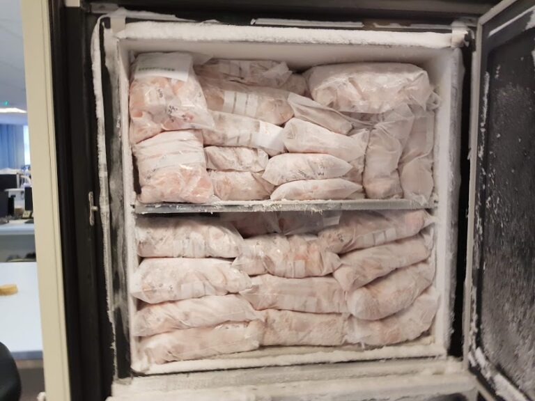
NAD+ (nicotinamide adenine dinucleotide, CAS 53-84-9) is one of the most widely studied coenzymes in biochemistry — and one of the most frequently mishandled reference materials on the research bench. The lyophilised white powder arrives in a vial looking perfectly inert; the chemistry that emerges the moment water is added is anything but. This guide works through the specific degradation mechanisms of NAD+ in solution, explains the critical pH asymmetry between the oxidised and reduced forms, and translates that chemistry into concrete handling decisions for a laboratory working with NAD+ strictly as a research-use-only reference material. Nothing here concerns human or veterinary use.
What NAD+ actually is, structurally — and where it breaks
NAD+ (nicotinamide adenine dinucleotide, C21H27N7O14P2, MW 663.43) is a dinucleotide assembled from two nucleotide moieties — adenosine monophosphate and nicotinamide mononucleotide — joined by a pyrophosphate bridge. The molecule carries two chemically distinct functional sites that govern its stability in solution:
The N-glycosidic bond between the nicotinamide ring and ribose is the dominant degradation site in the oxidised form. Under alkaline conditions, the hydroxide ion attacks the anomeric carbon, cleaving this bond and liberating free nicotinamide and adenosine diphosphoribose (ADPR) — a chemically inert non-redox-active product. This pathway accelerates approximately tenfold for each pH unit above neutral.12
The pyrophosphate bridge connecting the two nucleotide halves is more resistant under ordinary laboratory conditions but susceptible to non-specific phosphodiesterases and to prolonged high-temperature incubation. White (1984) studied the hydrolytic stability of NAD’s pyrophosphate and N-glycosidic bonds as part of a broader investigation of biomolecule stability at extreme temperatures, providing rate constants that frame what “stable” means under defined conditions.3
In the reduced form (NADH), an additional site opens up: the 1,4-dihydropyridine ring of the nicotinamide moiety. This ring is thermodynamically activated and susceptible to acid-catalysed decomposition at pH below 6, oxidation by dissolved oxygen, and UV photolysis at 340 nm. The important practical consequence is that NAD+ and NADH are not simply interchangeable forms of “the same molecule” from a handling perspective — they have opposite pH stability profiles.
2 opposite pH stability windows — NAD+ alkaline-labile above pH 7; NADH acid-labile below pH 6 — making buffer choice the most consequential single variable in a NAD+/NADH assay.
The differential stability of NAD+ and NADH: a mechanistic account
The pH asymmetry between NAD+ and NADH is documented in systematic stability studies and is the foundation for every practical handling decision. Rover Júnior et al. (1998) investigated the chemical stability of NADH/NAD+ and NADPH/NADP+ using UV-visible spectrophotometric monitoring across pH ranges and temperature conditions, establishing that buffer composition and pH are the dominant chemical determinants of cofactor degradation rate.1 Wu et al. (1986) specifically quantified the kinetics of NADPH degradation — the phosphorylated analog — reporting half-life data across pH and temperature ranges and demonstrating that effects of ionic strength (phosphate vs. acetate buffers) are secondary to the primary pH and temperature factors.2
The mechanistic picture for each form:
- NAD+ (oxidised, the form supplied as research powder): the nicotinamide ring is fully aromatic and electron-deficient, making the glycosidic bond susceptible to nucleophilic attack by hydroxide. Degradation above pH 7 is slow at 4 °C but becomes analytically significant at room temperature. The products (free nicotinamide, ADPR) are not redox-active and cannot substitute for intact NAD+ in enzymatic assays.
- NADH (reduced, generated in situ during enzymatic assays): the dihydropyridine ring is electron-rich and thermodynamically meta-stable. It decomposes in acid, is readily oxidised to NAD+ by dissolved oxygen, and is photolysed at 340 nm — the very wavelength used to monitor NADH formation in kinetic assays. Yin et al. (2026) demonstrated that NADH in reductase assay buffers degrades measurably within minutes at room temperature under standard conditions, and that imidazole (1–10 mM) significantly retards this decomposition, primarily through a pH-buffering and ring-stabilising effect.4
NAD+ is alkaline-labile; NADH is acid-labile and oxygen-labile. A buffer that suits one form degrades the other. Knowing which form is the assay substrate determines the correct pH strategy.
Temperature: the rate multiplier
Across both degradation profiles, temperature is the master variable. The Arrhenius relationship governs hydrolysis as it governs almost every chemical reaction: rate constants roughly double for each 10 °C increase in temperature. The practical implications are stark:
- NAD+ at −20 °C vs. 4 °C: approximately 10-fold difference in degradation rate.
- NAD+ at 4 °C vs. 22 °C (room temperature): another approximately 10-fold difference.
- Cumulative effect: a reconstituted NAD+ solution left on the bench at room temperature degrades roughly 100 times faster than the same solution on ice.
McDonough et al. (2025) quantified the thermal lability of NAD+ directly, reporting that the “poor thermal stability of the cofactor NAD+” in aqueous solution is a practical limitation that drives engineering solutions for industrial biosynthetic applications — documenting half-life data as a function of temperature that gives concrete numbers to the Arrhenius framework.5 Taniguchi et al. (2019) described “the problem of NAD+ instability at high temperatures” as the central obstacle for in vitro metabolic engineering at elevated temperatures, providing further quantitative context.6
Why the lyophilised state is so different from the dissolved state
The lyophilised powder does not resist degradation because it is chemically more stable than the dissolved molecule. It resists degradation because freeze-drying removes water — which is both the reactant and the medium for every hydrolysis pathway. Without water, the N-glycosidic bond cannot be cleaved; without water as a solvent, thermally driven oxidative reactions proceed at negligible rates. The dry amorphous solid is a kinetically arrested state, not a thermodynamically stable one.
This framing matters because it immediately explains the two most common ways researchers inadvertently degrade their NAD+ stock before any experiment begins. First: opening a cold vial in a warm, humid laboratory condenses atmospheric moisture onto the cold powder surface — the sealed vial was designed to prevent this, and bringing it to room temperature before unsealing eliminates the temperature differential that drives condensation. Second: a vial resealed imperfectly, or stored with a compromised septum, slowly equilibrates with ambient humidity, introducing the water that freeze-drying removed. The degradation in both cases is chemically identical to reconstitution — just uncontrolled and invisible.
Storage conditions — a decision table
| Variable | Recommended condition | Mechanism — why it matters | Risk if violated |
|---|---|---|---|
| Temperature (powder) | −20 °C (routine); −80 °C (long-term archive) | Arrhenius: hydrolysis rate ~2× per 10 °C rise; −20 °C gives ~100× slower rate than room temperature5 | Accelerated N-glycosidic hydrolysis; purity below COA specification |
| Temperature (reconstituted solution) | On ice (0–4 °C); use within 4–8 h or snap-freeze | Aqueous hydrolysis proceeds at all temperatures; refrigeration provides hours of stability, not days1 | Loss of functional NAD+; erroneous assay baselines; NADH oxidation drift |
| pH (NAD+ solution) | 6.0–7.0 for stock; 6.5–7.5 for assay buffer | Base-catalysed N-glycosidic hydrolysis accelerates ~10× per pH unit above neutral12 | Rapid glycosidic cleavage; loss of redox-active NAD+; free nicotinamide contaminates solution |
| pH (NADH solution) | 7.0–8.0; avoid <6 | Dihydropyridine ring is acid-labile; also oxidised by O₂ at all pH4 | Acid-catalysed ring decomposition; false-low NADH signal; overestimated enzyme activity |
| Humidity (powder) | Sealed, desiccated; equilibrate to room temperature before opening | Hygroscopic powder absorbs moisture from air; condensation on cold powder reintroduces water | Partial reactivation of aqueous hydrolysis in powder; clumping; silent purity loss |
| Light / UV | Amber vial or foil wrap; avoid direct UV and intense artificial light | NADH photolysed at 340 nm (dihydropyridine chromophore); NAD+ more resistant but not immune | NADH loss in assay solutions; interference at the 340 nm detection wavelength |
| Oxygen exposure | Minimise headspace in microtubes; use within working session | NADH oxidised back to NAD+ by dissolved O₂ continuously; bench-top NADH solutions deplete spontaneously4 | Underestimated NADH concentration; unexpected NAD+/NADH ratio; reductase assay drift |
| Freeze-thaw cycles | Aliquot before first freeze; ≤1 thaw per aliquot | Each cycle risks ice-crystal stress and pH transients at the freezing front5 | Cumulative degradation; inconsistent NAD+ concentrations across an experiment time course |
Storage and handling conditions for Condor Research lyophilised NAD+ (1000 mg vial, ≥99% HPLC, third-party tested in Czechia). All conditions describe laboratory sample preparation for in-vitro research only — not human or veterinary use. Consult the lot-specific COA for release specification data.
Reconstitution in practice: step by step
The following describes laboratory sample preparation for research use. It is not a protocol for any application to persons or animals.
- Remove the vial from −20 °C and allow it to reach room temperature (sealed). This step is not optional: cold powder in warm humid air condenses moisture that begins hydrolysis. Full equilibration typically takes 20–30 minutes.
- Prepare reconstitution buffer before opening the vial. For most enzyme assays: 50 mM sodium phosphate or HEPES at pH 6.5–7.5, prepared with ultrapure water (≥18 MΩ·cm). Confirm pH before use. Avoid buffers at pH >8 (accelerated base hydrolysis) and carbonate buffers. Tris-HCl at pH 7.0–7.5 is acceptable for most applications.
- Dissolve the powder rapidly and transfer to ice. Add a small volume of buffer directly to the vial, mix gently (invert or pipette), and place immediately on ice. Do not vortex vigorously.
- Aliquot before freezing. Divide the stock into single-experiment volumes in pre-chilled, light-protected microtubes. Label with date and lot number.
- Snap-freeze in liquid nitrogen and store at −20 °C or −80 °C. Snap-freezing minimises ice-crystal damage and pH transients. Use each aliquot once.
- Protect from light throughout. Work in subdued lighting; wrap microtubes in aluminium foil when not in active use.
The general principles of handling lyophilised research materials — the rationale for cold-chain discipline, moisture protection, and single-use aliquoting — are covered in the companion guide on how to store and reconstitute a lyophilised peptide. The pH and oxidation sensitivities specific to NAD+ described here are not shared by most peptide materials and require the additional considerations above.
Verifying solution integrity before an assay
A COA establishes what was in the vial at the point of manufacture. It cannot certify what is in the reconstituted solution on your bench. Pre-assay verification eliminates that uncertainty.
Spectrophotometric check: measure A260 of the stock solution and compare to a freshly prepared reference at the same nominal concentration. Intact NAD+ has a characteristic adenine absorption at 259–260 nm; degradation products (free nicotinamide, free adenine) shift and broaden this profile. For NADH stocks, the A340/A260 ratio should match the expected value — absent A340 in a supposedly reduced preparation means re-oxidation has occurred. The HILIC–MS/MS platform described by Røst et al. (2020) provides the highest-precision quantification of pyridine nucleotides in biological matrices, with sample handling protocols designed to prevent NAD+ degradation during extraction — a useful benchmark for laboratory stock-solution quality control.7
Enzymatic cycling check: the standard functional verification uses a cycling assay — typically lactate dehydrogenase for NAD+ or glucose-6-phosphate dehydrogenase for NADP+ — and compares the observed A340 change per unit time to the stoichiometric expectation from the gravimetric concentration. Gibon and Larher (1997) described the NaCl precipitation / ethanol solubilisation cycling protocol that remains a reference method for nicotinamide nucleotide quantification in plant and biochemical extracts; the same functional logic applies to stock-solution verification.8 A discrepancy between spectrophotometric and enzymatic assay values is the clearest indicator of degradation — the degradation products are visible to UV but inactive in the enzyme assay.
The research context: why NAD+ material quality matters
NAD+ is consumed — not merely used — by the signalling enzymes that make it interesting to study. Sirtuins cleave the glycosidic bond to release nicotinamide during deacylation; PARP enzymes consume NAD+ to synthesise poly(ADP-ribose) in response to DNA damage; CD38 hydrolyses NAD+ to cyclic ADP-ribose or ADPR as second messengers.9 In each case, the assay depends on the NAD+ supplied being fully intact. A partially degraded stock does not simply reduce the signal by the fraction lost — it introduces ADPR, free nicotinamide, or other products that may themselves act as enzyme inhibitors or substrates, producing a confounded result rather than a scaled-down clean one.
The Covarrubias, Perrone, Grozio and Verdin (2021) review in Nature Reviews Molecular Cell Biology provides the most authoritative current framework for NAD+ biology in the context of cellular ageing — summarising biosynthetic routes, consumer enzymes, and the mechanisms linking NAD+ decline to hallmarks of ageing that motivate much of the research interest in NAD+ as a reference material.9 The Migaud, Ziegler and Baur (2024) Nature Reviews Molecular Cell Biology overview updates the regulatory and therapeutic landscape.10 Both reviews assume, implicitly, that the NAD+ supplied in any assay is what it claims to be — an assumption that depends entirely on the handling practices described above.
All materials supplied by Condor Research are Research Use Only (RUO). Nothing in this guide constitutes guidance for human supplementation, clinical administration, or veterinary use. The mechanisms described are drawn from in-vitro and analytical biochemistry literature. In a study, the researchers reported — or an assay demonstrated — the pattern described; this does not establish efficacy or safety in any organism.
Condor Research NAD+ (product 351) is characterised at ≥99% purity by HPLC, confirmed by mass spectrometry, and tested at an independent EU laboratory in the Czech Republic. The COA is available on the NAD+ product page. This compound is supplied strictly as a Research Use Only reference material — not for human or veterinary use, not for diagnostic or therapeutic application. The chemistry summarised above is drawn from published analytical and biochemical literature and does not constitute a dosing protocol, clinical guidance, or safety assessment for any organism.
Condor Research · Scientific desk
Atrio Sciences s.r.o., IČO 57 669 651, Nitra (SK) · info@condorresearch.com
- NAD+ (oxidised form) is alkaline-labile: the N-glycosidic bond between nicotinamide and ribose hydrolyses above pH 7, producing free nicotinamide and ADP-ribose.
- NADH (reduced form) shows the inverse profile — stable in mild alkali, acid-labile below pH 6, and additionally vulnerable to oxidation by dissolved oxygen and UV photolysis at 340 nm.
- Temperature is the master stability variable: hydrolysis rate roughly doubles per 10 °C (Arrhenius), making the difference between −20 °C storage and room temperature analytically decisive.
- Lyophilised powder is far more stable than aqueous solution because freeze-drying removes water — the reactant and medium for all degradation routes.
- Opening a cold vial in ambient air condenses moisture onto the powder, restarting aqueous degradation silently. Equilibrate the sealed vial to room temperature before opening.
- Reconstituted solutions should be prepared on ice, protected from light, and used within 4–8 h; aliquot into single-use volumes before first freeze.
- A COA certifies purity at release; it does not certify the material after reconstitution. Verify solution integrity at A₂₆₀ before critical assays.
How long does lyophilised NAD+ remain stable at −20 °C?
Under proper conditions — sealed, desiccated, −20 °C, protected from light — lyophilised NAD+ is expected to remain within specification for at least 12–24 months as a research reference material. Exact shelf-life is lot-specific and stated on the Certificate of Analysis supplied with each vial. Do not extrapolate shelf-life data from one lot to another without current COA documentation.
Can I store a reconstituted NAD+ solution in the fridge overnight?
Short-term refrigeration at 4 °C in a sealed, light-protected tube at pH 7.0–7.5 for up to 24 hours is reported in published enzymatic protocols and is generally adequate for applications where absolute NAD+ concentration is not a critical variable. Where it is critical — kinetic rate studies, sirtuin activity assays with NAD+ as the limiting substrate — the recommended practice is to snap-freeze single-use aliquots immediately after preparation and thaw only once. Measurable degradation does occur over a 24-hour refrigerated window at neutral pH.
Why does NAD+ degrade faster in alkaline buffer than in neutral buffer?
The N-glycosidic bond linking nicotinamide to ribose is susceptible to base-catalysed hydrolysis. Hydroxide ion (OH⁻) concentrations increase tenfold per pH unit above neutral (pH 7), attacking the anomeric carbon of the nicotinamide-ribose linkage. Above pH 8, the rate of cleavage is fast enough to be analytically significant within minutes at room temperature. The products — free nicotinamide and ADPR — are not redox-active and cannot substitute for intact NAD+ in enzyme assays.
What reconstitution solvent is best for NAD+ enzyme assays?
Phosphate (50 mM, pH 6.5–7.5) or HEPES (25–50 mM, pH 6.5–7.5) buffer in ultrapure water is standard for most NAD+-dependent enzyme assays. Tris-HCl at pH 7.0–7.5 is widely used and acceptable. Avoid alkaline buffers (pH > 8 accelerates glycosidic hydrolysis), carbonate buffers, and DMSO for NAD+ stocks. For applications where NADH must be maintained in a stable reduced state during an extended kinetic read at room temperature, adding imidazole (1–10 mM) to the assay buffer has been shown to retard dihydropyridine ring decomposition.
How do I know if my reconstituted NAD+ has degraded?
Spectrophotometrically: compare A260 against a freshly prepared reference solution at the same gravimetric concentration. Degradation products shift and broaden the UV spectrum. Enzymatically: run an LDH-coupled assay and confirm that the A340 rise per micromole of NAD+ added matches the theoretical stoichiometry. A discrepancy between spectrophotometric and enzymatic values — where spectrophotometry indicates full concentration but enzymatic assay shows reduced activity — confirms the presence of spectrophotometrically active but enzymically inert degradation products.
Does the Condor Research NAD+ vial need reconstitution before use?
For solution-phase enzymatic assays, binding studies, cell-free systems, and metabolite quantification: yes, aqueous reconstitution is required. For use as a dry reference standard in HPLC or MS calibration: the powder can be weighed directly and dissolved gravimetrically in the appropriate analytical solvent. Consult the lot-specific COA for residual moisture data, which is needed for accurate gravimetric calculations.